Dinokyste
Un dinokyste ou kyste de dinoflagellé constitue le stade dormant et zygotique du cycle de vie de certains dinoflagellés. Ils ont généralement un diamètre de 15 à 100 µm et sont produits par environ 15 à 20 % des dinoflagellés vivants.
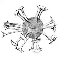
Ils peuvent s'accumuler dans les sédiments sous forme de microfossiles. Les dinocystes à parois organiques sont souvent résistants et constitué de dinosporine (en). Il existe également des kystes de dinoflagellés calcaires et d'autres siliceux. De nombreux ouvrages décrivent les dinocystes[1].
Historique
La première personne à reconnaitre les dinoflagellés fossiles fut Christian Gottfried Ehrenberg, qui a rapporté sa découverte dans un article présenté à l'Académie des sciences de Berlin en . Il avait observé des dinoflagellés clairement tabulés dans de minces flocons de silex du Crétacé et considérait ces dinoflagellés comme ayant été silicifiés. Avec eux, se trouvaient des corps sphéroïdaux à ovoïdes de taille comparable et portant un ensemble de protubérances ou de tubes de formes diverses. Ehrenberg les a interprétés comme étant à l'origine siliceux et y a vu des Desmidiées (des algues d'eau douce) et les a placés dans le genre Xanthidium (en) qu'il venait de créer. Bien que des résumés les travaux d'Ehrenberg aient paru plus tôt, les études n'ont été publiés dans leur intégralité que vers 1837 ou 1838[2].
Un premier lien entre les thèques et kystes de dinoflagellés a été établi par leur comparaison morphologique qu'ont effectuée Bill Evitt et Susan E. Davidson[3]. D'autres preuves ont été établies par des études détaillées de culture de dinokystes menées par David Wall et Barrie Dale à la Woods Hole Oceanographic Institution dans les années 1960[4],[5].
Typologie
Sur le plan ontologique, le terme kyste peut s'appliquer à :
- un état de repos temporaire (kyste pelliculaire, temporaire ou ecdysal) ;
- un zygote dormant (kyste au repos ou hypnozygote) ;
- un état coccoïde dans lequel les cellules restent photosynthétiquement actives[6].
Par exemple pour ce dernier cas particulier, tous les kystes produits par des espèces de l'ordre des Phytodiniales (genres Cystodinium, Stylodinium, Hypnodinium, Tetradinium, Dinococcus, Gloeodinium) sont des stades coccoïdes.
Un kystes digestif désigne un kyste pelliculaire formé après phagocytose comme chez Katodinium fongiforme[7],[8].
Un kyste de division se réfère à un stade non mobile dans lequel la reproduction asexuée s'effectue par division[9]. Ce ne sont pas des kystes pelliculaires ou au repos car ils ne sont pas dormants. De même, les stades palmelloïdes ou mucilage ne sont pas des kystes pelliculaires ou au repos, mais des stades au cours desquels la monade perd ses flagelles et s'enveloppe de plusieurs couches de mucilage pour effectuer sa division[10].
Taxonomie
Les dinokystes décrits dans la littérature sont liés à un stade mobile particulier par des similitudes morphologiques, une cooccurrence dans la même population/culture Ces relations peuvent être établies par la technique d'incubation des kystes[11],[5],[12],[13]. Les géologues utilisent une taxonomie basée sur les kystes, tandis que les biologistes utilisent une taxonomie basée sur les stades mobiles. Par conséquent, les kystes peuvent avoir des noms différents de ceux de leur stades mobile correspondant. Les kystes vivants peuvent être facilement dissociés des sédiments au moyen de polytungstate de sodium, un liquide de forte densité[14]. Une autre méthode, rarement utilisée, utilise un gradient de saccharose[15]. Plus récemment, on a pu identifier des séquences moléculaires de kystes et de cellules[16],[17],[18].
15 à 20 % les dinoflagellés marins[19] et 24 % des dinoflagellés d'eau douce[20] forment des kystes. La tabulation du dinoflagellé se reflète parfois dans la tabulation (autrefois appelée paratabulation) de son dinokyste, permettant ainsi de déduire l’espèce à partir du kyste[21]. Il a déjà été suggéré que, chez les espèces marines, les caractères morphologiques du stade kystique seraient significatifs d'un point de vue phylogénétique[22] et cela serait encore plus vrai pour les dinoflagellés d'eau douce[23], confirmés par de nouvelles observations[24],[25] et récemment révisé[20]. Plusieurs livres documentent la taxonomie générale des kystes[21],[26]. Il existe peu de guides pour la détermination des dinokystes marins du Quaternaire[27],[28]. De nombreuses espèces nouvelles sont encore en cours de description pour le Néogène[29] (qui couvre le Miocène[30],[31] et le Pliocène[32],[33],[34],[35]) et le Quaternaire (qui couvre le Pléistocène[36] et l'holocène[37],[38],[39]).
Description
Taille
Les dinokystes quaternaires ont généralement un diamètre compris entre 15 et 100 µm[40]. L'un des plus petits kystes récents est le kyste de Pentapharsodinium dalei, qui peut mesurer jusqu'à 19 µm de long[41]. L'un des plus gros kystes récents est le kyste de Protoperidinium latissimum, qui peut mesurer jusqu'à 100 µm de long[5].
Composition
Les parois des dinocystes à parois organiques sont composées d'un biopolymère résistant appelé dinosporine[42]. Ce composé organique a des similitudes avec la sporopollénine, mais est spécifique aux dinoflagellés.
En plus des kystes à parois organiques, il existe également des dinokystes calcaires et d'autres siliceux.
Ultrastructure du kyste
Il y a peu d'études ultrastructurales de kystes marins en MET, à l'exception de Hystrichosphaea bentorii, Hystrichosphaeridium, Impletosphaeridium, Lingulodinium machaerophorum, Operculodinium centrocarpum, Bitectatodinium tepikiense[43],[44],[45] et plus récemments sur Lingulodinium machaerophorum[46] et Alexandrium[47].
Certains kystes d'eau douce ont été étudiés par MET, comme Ceratium hirundinella[48].
Cycle de vie
Les kystes au repos sont une étape du cycle sexuel des Dinoflagellés[49]. Induite par des déclencheurs particuliers tels que des changements de température, de nutriments[50], etc., la formation de gamètes intervient. Les gamètes fusionnent pour former le planozygote et subissent un enkystement : ils forment des kystes au sein des thèques du planozygote. Ceux-ci tombent rapidement dans les sédiments. De nombreuses espèces peuvent passer de plus longues périodes de repos dans les sédiments que d'activité dans la colonne d'eau[51]. Les stades de repos constituent également un réservoir de diversité génétique, ce qui augmente le potentiel de survie des populations[52]. Ainsi, les kystes de dinoflagellés ont une grande importance écologique et agissent comme des « banques de graines », comparables à celles que l'on trouve dans les écosystèmes terrestres. Les formes enkystées peuvent rester viables jusqu'à 100 ans[53]. Les sédiments peuvent stocker des kystes de Lingulodinium vivants pendant au moins 18 mois[54]. Les kystes ont souvent besoin de déclencheurs pour germer, tels que des changements de température, de nutriments, etc. Certains kystes, tels que Scrippsiella acuminata, nécessitent de la lumière pour germer[55].
Notes et références
- (en) Cet article est partiellement ou en totalité issu de l’article de Wikipédia en anglais intitulé « Dinocyst » (voir la liste des auteurs).
- Evitt, W. R. 1985. Sporopollenin Dinoflagellate Cysts: Their Morphology and Interpretation. American Association Stratigraphic Palynologists Monograph Ser. 1.
- W.A.S. Sarjeant, 2002. 'As chimney-sweeps, come to dust': a history of palynology to 1970. pp. 273–327 In: Oldroyd, D. R. The earth inside and out: some major contributions to geology in the twentieth century. Geological Society (London) Special Publication no. 192.
- Evitt, W.R. and Davidson, S.E. 1964. Dinoflagellate studies. 1. Dinoflagellate cysts and thecae. Stanford university publications X (1), pp. 3–12.
- D. Wall et B. Dale, « Living" fossils in western Atlantic plankton », Nature, vol. 211, no 5053, , p. 1025–1026 (DOI 10.1038/2111025a0, Bibcode 1966Natur.211.1025W)
- D. Wall et B. Dale, « Modern dinoflagellate cysts and evolution of the Peridiniales », Micropaleontology, vol. 14, no 3, , p. 265–304 (DOI 10.2307/1484690, JSTOR 1484690)
- Pfiester L.A. & Anderson D.M. 1987. Dinoflagellate reproduction. In: The biology of dinoflagellates. Botanical monographs 21 (Ed. by F.J.R. Taylor), pp. 611–648., Blackwell Scientific Publications.
- W.A.S. Sarjeant, T. Lacalli et G. Gaines, « The cysts and skeletal elements of dinoflagellates: speculations on the ecological causes for their morphology and development », Micropaleontology, vol. 33, no 1, , p. 1–36 (DOI 10.2307/1485525, JSTOR 1485525)
- H.J. Spero et M.D. Moree, « Phagotrophic feeding and its importance to the life cycle of the holozoic dinoflagellate Gymnodinium fungiforme », Journal of Phycology, vol. 17, , p. 43–51 (DOI 10.1111/j.1529-8817.1981.tb00817.x)
- Bravo I., Figueroa R.I., Garcés E., Fraga S. & Massanet A. 2010. The intricacies of dinoflagellate pellicle cysts: the example of Alexandrium minutum cysts from a bloom-recurrent area (Bay of Baiona, NW Spain). Deep-Sea Research Part II: Topical Studies in Oceanography 57: 166–174.
- Popovský J. & Pfiester L.A. 1990. Dinophyceae (Dinoflagellida). In: Süßwasserflora von Mitteleuropa. Begründet von A. Pascher. Band 6 (Ed. by H. Ettl,J. Gerloff,H. Heynig. & D. Mollenhauer). Gustav Fischer Verlag, Jena, 272 pp.
- D. Wall et B. Dale, « Living fossils" in Atlantic plankton », Nature, vol. 211, no 5053, , p. 1025–1026 (DOI 10.1038/2111025a0)
- J.A. Sonneman et D.R.A. Hill, « A taxonomic survey of cyst-producing dinoflagellates from recent sediments of Victorian coastal waters, Australia », Botanica Marina, vol. 40, nos 1–6, , p. 149–177 (DOI 10.1515/botm.1997.40.1-6.149)
- K.N. Mertens, A. Yamaguchi, H. Kawami, S. Ribeiro, B.S. Leander, A.M. Price, V. Pospelova, M. Ellegaard et K. Matsuoka, « Archaeperidinium saanichi sp. nov.: a new species based on morphological variation of cyst and theca within the Archaeperidinium minutum Jörgensen 1912 species complex », Marine Micropaleontology, vol. 96–97, , p. 48–62 (DOI 10.1016/j.marmicro.2012.08.002, Bibcode 2012MarMP..96...48M)
- C.J.S. Bolch, « The use of polytungstate for the separation and concentration of living dinoflagellate cysts from marine sediments », Phycologia, vol. 36, no 6, , p. 472–478 (DOI 10.2216/i0031-8884-36-6-472.1)
- P. Schwinghamer, D.M. Anderson et D.M. Kulis, « Separation and concentration of living dinoflagellate resting cysts from marine sediments via density-gradient centrifugation; », Limnology and Oceanography, vol. 36, no 3, , p. 588–592 (DOI 10.4319/lo.1991.36.3.0588, Bibcode 1991LimOc..36..588S)
- C.J.S. Bolch, « PCR protocols for genetic identification of dinoflagellates directly from single cysts and plankton cells », Phycologia, vol. 40, no 2, , p. 162–167 (DOI 10.2216/i0031-8884-40-2-162.1)
- Y. Takano et T. Horiguchi, « Acquiring scanning electron microscopical, light microscopical and multiple gene sequence data from a single dinoflagellate cell », Journal of Phycology, vol. 42, , p. 251–256 (DOI 10.1111/j.1529-8817.2006.00177.x)
- H. Kawami, R. Van Wezel, R.P. Koeman et K. Matsuoka, « Protoperidinium tricingulatum sp. nov. (Dinophyceae), a new motile form of a round, brown, and spiny dinoflagellate cyst », Phycological Research, vol. 57, no 4, , p. 259–267 (DOI 10.1111/j.1440-1835.2009.00545.x)
- HEAD M.J. 1996. Modern dinoflagellate cysts and their biological affinities. In: Palynology: principles and applications (Ed. by J. Jansonius & D. C. McGregor), pp. 1197–1248. American Association of Stratigraphic Palynologists Foundation, Dallas, Texas.
- Kenneth Neil Mertens, Karin Rengefors, Øjvind Moestrup et Marianne Ellegaard, « A review of recent freshwater dinoflagellate cysts: taxonomy, phylogeny, ecology and palaeocology », Phycologia, vol. 51, no 6, , p. 612–619 (DOI 10.2216/11-89.1)
- R.A. Fensome, F.J.R. Taylor, G. Norris, W.A.S. Sarjeant, D.I. Wharton et G.L. Williams, « A classification of living and fossil dinoflagellates », American Museum of Natural History, Micropaleontology, Special Publication, vol. 7, , p. 1–351
- R Harland, « A review of Recent and Quaternary organic-walled dinoflagellate cysts of the genus Protoperidinium », Palaeontology, vol. 25, , p. 369–397
- A.J. Schilling, « Die Süsswasser-Peridineen », Flora Oder Allgemeine Botanische Zeitung, vol. 74, , p. 220–299
- M. Tardio, M. Ellegaard, N. Lundholm, F. Sangiorgi et D. DI Giuseppe, « A hypocystal archeopyle in a freshwater dinoflagellate from the Peridinium umbonatum group (Dinophyceae) from Lake Nero di Cornisello, South-East Alps, Italy », European Journal of Phycology, vol. 44, no 2, , p. 1–10 (DOI 10.1080/09670260802588442)
- Ø. Moestrup, K. Lindberg et N. Daugbjerg, « Studies on woloszynskioid dinoflagellates IV: The genus Biecheleria gen. nov », Phycological Research, vol. 57, no 3, , p. 203–220 (DOI 10.1111/j.1440-1835.2009.00540.x)
- Evitt, W.R., Lentin, J.K., Millioud, M.E., Stover, L.E. and Williams, G.L., 1977. Dinoflagellate cyst terminology. Geological survey of Canada, Paper 76-24, 1-11.
- Rochon, A., de Vernal, A., Turon, J.-L., Matthiessen, J., and Head, M.J., 1999. Distribution of recent dinoflagellate cysts in surface sediments from the North Atlantic Ocean and adjacent seas in relation to sea-surface parameters. AASP Contribution Series, 35, 146 pp.
- MATSUOKA, K. & FUKUYO, Y. 2000. Technical guide for modern dinoflagellate cyst study. WESTPAC-HAB/WESTPAC/IOC, Japan Society of the Promotion Science, Tokyo, 29 pp.
- M.J. Head et G. Norris, « New species of dinoflagellate cysts and other palynomorphs from the late Neogene of the western North Atlantic, DSDP Hole 603C », Journal of Paleontology, vol. 77, , p. 1–15 (DOI 10.1666/0022-3360(2003)077<0001:nsodca>2.0.co;2)
- S. Louwye, K.N. Mertens et D. Vercauteren, « New dinoflagellate cysts species from the Miocene of Porcupine Basin, off Southwest Ireland », Palynology, vol. 32, , p. 131–142 (DOI 10.2113/gspalynol.32.1.131)
- Soliman, A., Head, M.J., and Louwye, S. In press. Morphology and distribution of the Miocene dinoflagellate cyst Operculodinium? borgerholtense Louwye 2001, emend. Palynology.
- Head, M.J., 1999. The Late Pliocene St. Erth Beds of Cornwall: a review of the palynology and reappraisal of the dinoflagellates. In: Scource, J. and Furze, M.F.A. (eds.), The Quaternary of West Cornwall. Field Guide, Quaternary Research Association, Durham, U.K., p. 88–92.
- Head, M.J. 2000. Geonettia waltonensis, a new goniodomacean dinoflagellate from the Pliocene of the North Atlantic region, and its evolutionary implications" Journal of Paleontology 74(5): 812–827, 6 pls.
- S. De Schepper, M.J. Head et S. Louwye, « New dinoflagellate cyst and incertae sedis taxa from the Pliocene of northern Belgium, southern North Sea Basin », Journal of Paleontology, vol. 78, no 4, , p. 625–644 (DOI 10.1666/0022-3360(2004)078<0625:ndcais>2.0.co;2)
- S. De Schepper et M.J. Head, « New dinoflagellate cyst and acritarch taxa from the Pliocene and Pleistocene of the eastern North Atlantic (DSDP Site 610) », Journal of Systematic Palaeontology, vol. 6, , p. 101–117 (DOI 10.1017/s1477201907002167)
- M.J. Head, « Echinidinium zonneveldiae sp. nov., a new dinoflagellate cyst from the Late Pleistocene of the Baltic region », Journal of Micropalaeontology, vol. 21, no 2, , p. 169–173 (DOI 10.1144/jm.21.2.169)
- T. Verleye, V. Pospelova, K.N. Mertens et S. Louwye, « The geographical distribution and (palaeo)ecology of Selenopemphix undulata sp. nov., a new late Quaternary dinoflagellate cyst from the Pacific Ocean », Marine Micropaleontology, vol. 78, nos 3–4, , p. 65–83 (DOI 10.1016/j.marmicro.2010.10.001, Bibcode 2011MarMP..78...65V)
- V. Pospelova et M.J. Head, « Islandinium brevispinosum sp. nov. (Dinoflagellata), a new organic-walled dinoflagellate cyst from modern estuarine sediments of New England (USA) », Journal of Phycology, vol. 38, no 3, , p. 593–601 (DOI 10.1046/j.1529-8817.2002.01206.x)
- K.N. Mertens, A. Yamaguchi, H. Kawami, S. Ribeiro, B.S. Leander, A.M. Price, V. Pospelova, M. Ellegaard et K. Matsuoka, « Archaeperidinium saanichi sp. nov.: a new species based on morphological variation of cyst and theca within the Archaeperidinium minutum Jörgensen 1912 species complex », Marine Micropaleontology, vol. 96–97, , p. 48–62 (DOI 10.1016/j.marmicro.2012.08.002, Bibcode 2012MarMP..96...48M)
- A. De Vernal et F. Marret, Organic-Walled Dinoflagellate Cysts: Tracers of Sea-Surface Conditions, vol. 1, , 371–408 p. (ISBN 9780444527554, DOI 10.1016/S1572-5480(07)01014-7)
- B Dale, « New observations on Peridinium faeroense Paulsen (1905), and classification of small orthoperidinioid dinoflagellates », Br. Phycol. J., vol. 12, no 3, , p. 241–253 (DOI 10.1080/00071617700650261)
- Fensome, R.A., Taylor, F.J.R., Norris, G., Sarjeant, W.A.S., Wharton, D.I., and Williams, G.L., 1993. A classification of modern and fossil dinoflagellates, Sheridan Press, Hanover.
- U Jux, « Über den feinbau der wandungen einiger Tertiärer Dinophyceen-zysten und Acritarcha Hystrichosphaeridium, Impletosphaeridium, Lingulodinium », Palaeontographica, Abt. B, vol. 132, nos 5–6, , p. 165–174
- U Jux, « Über den feinbau der wandungen bei Operculodinium centrocarpum (Deflandre & Cookson) Wall 1967 und Bitectatodinium tepikiense Wilson 1973 », Palaeontographica, Abt. B, vol. 155, nos 5–6, , p. 149–156
- J. P. Kokinos, T.I. Eglinton, M.A. Goñi, J.J. Boon, P.A. Martoglio et D.M. Anderson, « Characterisation of a highly resistant biomacromolecular material in the cellwall of a marine dinoflagellate resting cyst », Organic Geochemistry, vol. 28, no 5, , p. 265–288 (DOI 10.1016/s0146-6380(97)00134-4)
- Gay Kennaway et Jane Lewis, « An ultrastructural study of hypnozygotes of Alexandrium species (Dinophyceae) », Phycologia, vol. 43, no 4, , p. 355–363 (DOI 10.2216/i0031-8884-43-4-355.1)
- D. V. Chapman, J. D. Dodge et S. I. Heaney, « Cyst Formation in the Freshwater Dinoflagellaie Ceratium Hirundinella (Dinophyceae) », Journal of Phycology, vol. 18, , p. 121–129 (DOI 10.1111/j.1529-8817.1982.tb03165.x)
- H.A. VON Stosch, « Sexualität bei Ceratium cornutum (Dinophyta) », Die Naturwissenschaften, vol. 52, no 5, , p. 112–113 (DOI 10.1007/bf00626331)
- Pfiester, L. A. & Anderson, D. M. 1987. Dinoflagellate reproduction. In: The biology of dinoflagellates (ed. F. J. R. Taylor), pp. 611–648. - Blackwell, Oxford.
- RENGEFORS K. 1998. Seasonal succession of dinoflagellates coupled to the benthic cyst dynamics in Lake Erken, Sweden. Archiv für Hydrobiologie, Special Issues, Advances in Limnology 51: 123–141.
- T.J. Alpermann, B. Beszteri, U. John, U. Tillmann et A.D. Cembella, « Implications of life history transitions on the population genetic structure of the toxigenic marine dinoflagellate Alexandrium tamarense », Molecular Ecology, vol. 18, no 10, , p. 2122–2133 (PMID 19389181, DOI 10.1111/j.1365-294x.2009.04165.x)
- S. Ribeiro, T. Berge, N. Lundholm, T.J. Andersen, F. Abrantes et M. Ellegaard, « Phytoplankton growth after a century of dormancy illuminates past resilience to catastrophic darkness », Nature Communications, vol. 2, , p. 311 (PMID 21587228, PMCID 3113231, DOI 10.1038/ncomms1314, Bibcode 2011NatCo...2..311R)
- Jane Lewis, A. Harris, K. Jones et R. Edmonds, « Long-term survival of marine planktonic diatoms and dinoflagellates in stored sediment samples », Journal of Plankton Research, vol. 21, no 2, , p. 343–354 (DOI 10.1093/plankt/21.2.343)
- B. J. Binder et D. M. Anderson, « Green light-mediated photomorphogenesis in a dinoflagellate resting cyst », Nature, vol. 322, no 6080, , p. 659–661 (DOI 10.1038/322659a0, Bibcode 1986Natur.322..659B)
 Portail de la microbiologie
  Portail de la biologie marine
  Portail de la phycologie